01 / MECHANICAL
Acceleromyography
16 triaxial sensors
Sixteen AIS2IHTR triaxial accelerometers capture local mechanical signals at four sites on the left leg.


SAME-LIMB · OPEN RESEARCH DATASET
HUMIT Lab · Harbin Institute of Technology, Shenzhen
Synchronized acceleromyography, surface electromyography, and optical motion capture from 30 participants across 16 lower-limb task conditions.
Drag horizontally to rotate · Arrow keys when focused
Illustrative walking · Arms crossed over the chest
Physical-scale layout · Selected sensor enlarged for inspection
THE DATASET
SAME-Limb brings together acceleromyography, surface electromyography, and optical motion capture from everyday lower-limb training—a reproducible foundation for studying muscle activity and estimating joint angles.
Sixteen AIS2IHTR triaxial accelerometers capture local mechanical signals at four sites on the left leg.
Four LE230 surface EMG channels measure electrical activity at the matched RF, VM, TA, and GA muscle sites.
16 infrared cameras track 15 markers to provide reference angles for both knees and ankles.
ACQUISITION PROTOCOL
Eleven functional actions and five walking speeds. Select an activity for its essential instructions.
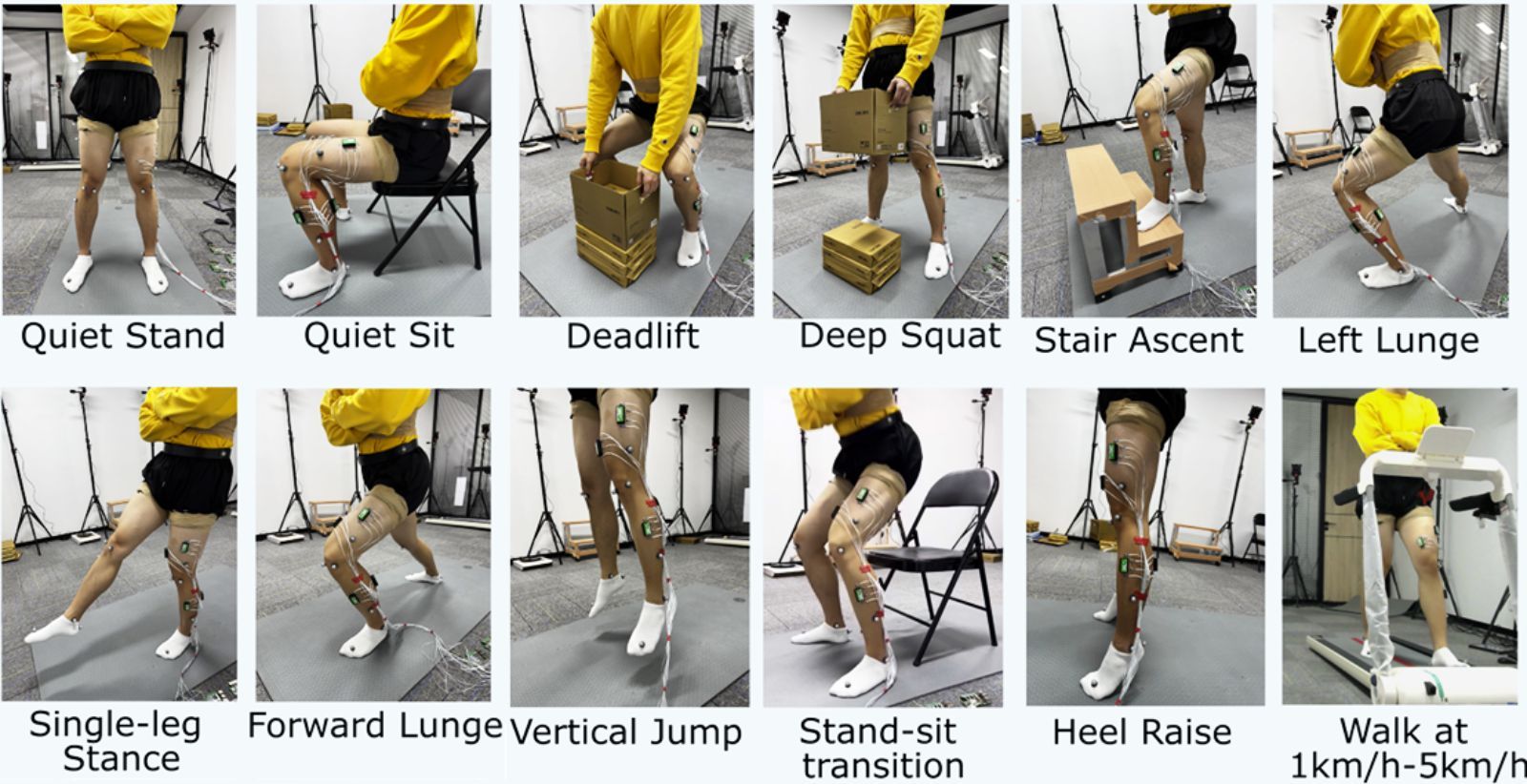
Use the same foot placement and height-adjusted box setup as the deadlift, with no standardized load inside. Complete the squat to the auditory cues.
Auditory cues at approximately 2, 6, and 10 s.REPRODUCIBLE BY DESIGN
Compare model performance, task-specific errors, and acquisition configurations using the published source data.
Table V · Pooled test-window metrics
Seed 42; one training run per configuration. Metrics are computed over valid pooled test windows for each of the four joint outputs, then averaged across outputs.
| Configuration | MAE (°) | RMSE (°) | Pearson r |
|---|---|---|---|
| Random Forest | 9.591 | 13.639 | 0.634 |
| TCN | 8.840 | 12.515 | 0.672 |
| LSTMPlus | 9.535 | 13.589 | 0.630 |
| InceptionTimePlus | 9.426 | 12.932 | 0.658 |
FROM SIGNAL TO MOVEMENT
Representative deep-squat, vertical-jump, and 1 km/h walking trials. Black traces show MoCap labels, dashed blue traces Random Forest, and pink traces TCN.
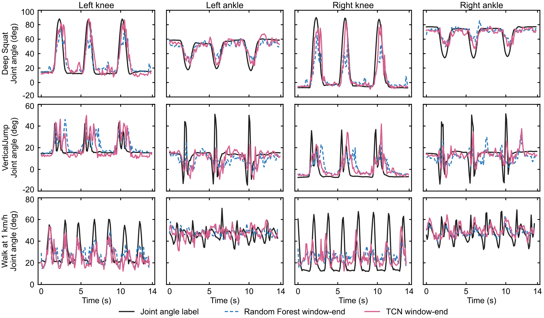 Fig. 4 · Open original
 Representative trials complement the aggregate evaluation; they do not replace it. Source: arXiv:2608.11958v1, Fig. 4.
READY FOR YOUR RESEARCH
1,918 NPZ files with AMG, EMG, MoCap, joint angles, validity masks, and modality-specific time vectors.
Browse trialsFixed splits, window manifests, 64 fitted model artifacts, reference predictions, and source tables.
Explore benchmark codeAMG streams share a common grid. All modalities are cropped to their shared interval; labels use the nearest native MoCap frame.
Read the data guideThe four AMG streams are linearly interpolated onto a nominal 1,600 Hz grid, without extrapolation. EMG retains its native 1,000 Hz sampling rate; MoCap retains native frame timestamps. Device clocks were coordinated by NTP, with no additional correction for cross-device clock offset or drift. Hardware-level synchronization accuracy was not established.
NPZ arrays include amg_raw_aligned, amg_dc_corrected_aligned, amg_highpass_5hz_aligned, semg_raw, semg_preprocessed_basic, motion_markers_named, motion_skeleton_selected, joint_angles_deg and joint_angle_valid_mask, provided alongside modality-specific time vectors and metadata_json. Raw AMG values are stored in g/digit; multiply by 19.14258 to convert to mm/s².
Auditory cue records are available for 1,726 trials and absent for 192. They were not used for alignment or benchmark computation.
Participants were healthy adults aged 23.4 ± 2.1 years. Clinical generalization to patients or other populations has not been validated. The study was approved by the Medical Ethics Committee of Harbin Institute of Technology (HIT-2024046; 8 July 2024). All participants provided written informed consent.
Data, metadata, research source materials, predictions, metrics, and trained models are licensed under CC BY 4.0; code/ software uses Apache-2.0. Third-party materials retain their original terms. The article and Supplementary Information have separate publication licenses.
The model uses a CC0 MakeHuman-derived mesh with an illustrative walking cycle and arms crossed over the chest. Transparent left-leg skin reveals schematic RF, VM, TA, and GA muscles. Fifteen reflective markers follow the lower-body segments; positions are schematic rather than measured participant coordinates.
LE230 geometry follows Biometrics dimensions: 42 × 24 × 14 mm. The AIS2IHTR IC measures 2 × 2 × 0.93 mm; its custom carrier board is illustrated at approximately 6 × 5 mm, estimated from paper Fig. 1. Devices use these dimensions in the physical-scale layout; selection temporarily enlarges one sensor for inspection.
Product reference images belong to Biometrics Ltd and STMicroelectronics. The human mesh was distributed through MakeHuman / MPFB2, NAVER anny, and three.ws.
View model license and hardware sources ↗THE PAPER
School of Computer Science and Technology
Harbin Institute of Technology, Shenzhen
@misc{tang2026synchronizedamgemgdataset,
      title={Synchronized AMG and EMG Dataset of Lower-limb Muscle Activities in Everyday Training},
      author={Dongxu Tang and Shih Ying-Lei and Zhuoyi Ren and Jianting Liao and Yitian Shao},
      year={2026},
      eprint={2608.11958},
      archivePrefix={arXiv},
      primaryClass={cs.HC},
      url={https://arxiv.org/abs/2608.11958},
}